

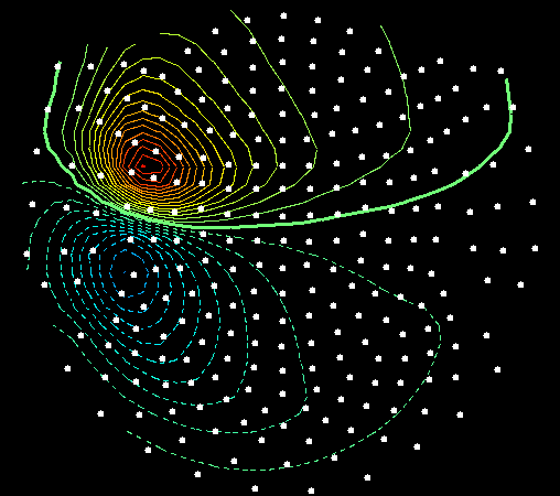
MEG ‘topographic’ map showing the distribution of radial magnetic field amplitudes produced by some underlying brain activity. In this case the contour lines represent areas of the same field intensity rather than areas of similar elevation. The two ‘peaks’ (the red and blue circles) can be used to estimate where the underlying activity was located. If you draw a line between the two peak centers, the activity is located below the midpoint of the line at a depth that is proportional to the distance between the centers.
Magnetoencephalography (MEG) is a non-invasive neuroimaging technique that maps functional brain activity by recording magnetic fields naturally produced by the brain’s electric activity. MEG has very high temporal resolution, and can record events on the order of milliseconds. Additionally, MEG has superior spatial resolution, and can localize sources with millimeter precision. Unlike fMRI or PET scans that are secondary measures, MEG directly measures brain function and neuronal activity.
How Does It Work?
Neurons use electric currents to communicate and facilitate activity in the brain. Electromagnetic fields are generated by the ionic current flowing through neurons. While the magnetic field generated by a single neuron is negligible, multiple neurons in a single area that are activated together generates a sizable magnetic field that can be recorded outside of the brain.
Although this neural activity can be measured, it is still approximately a billionth of the strength of Earth’s own magnetic field, and thus sensitive instruments are required to record brain activity. An array of superconducting quantum interference devices (or SQUIDS) that are cooled with liquid Helium to -269 degrees Celsius are able to detect and amplify the magnetic signals generated by neurons centimeters away. Recordings are performed in a magnetically shielded room that minimizes the amount of background magnetic interference.